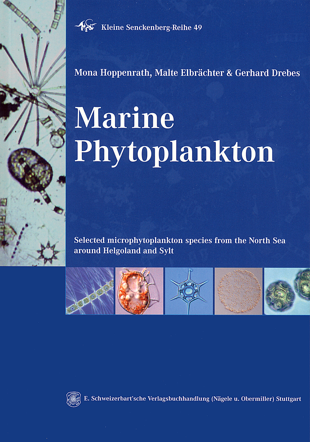
For many of us working with marine phytoplankton in the decades
following 1974 when it was published, Marines Phytoplankton by Gerhard
Drebes was a little gem. A compact volume in the series of pocketbooks
published by Thieme, it was a mystery why it was never reprinted and
translated from the German, particularly as it was always difficult to
keep one’s eyes on the copy because of it’s size and usefulness. Now,
at long last, we have its child, which in the way of things, is larger
and in English.
For this reviewer the key to success of the book will be the fact that
it will be excellent for students examining live samples of marine
phytoplankton in marine labs around the world even though it
concentrates on taxa that occur in samples from a comparatively small
area – the German Bight. It concentrates on the diatoms and
dinoflagellates; too small to be seen with the naked eye but large
enough to be distinguished in the light microscope. The great bulk of
the literature on the subject including texts available to students in
those laboratories, have largely lacked good illustrations of the
living cells. Now, with this book, there will be opportunity for lab
and class organizers to appeal to the general interest of young people
in organisms that move and do things, and this for two reasons. The
form of cell and colony, division cycles and sexual stages and even
infestations are covered for many species and at the end of the book
there is a long list of the beautiful films of Drebes and his
co-workers available now in DVD form at: www.iwf.de.
Comparison of the introductory sections of the two books shows how new
concerns have arisen in the past 35 years, viz. harmful and toxic taxa
and the changing face of the assemblages themselves due to invasions
of taxa tolerant of a changed environment or brought in by ballast
water from ships. The introduction of the present book and the
beautiful satellite image in the frontispiece tells us that the book
is devoted to the phytoplankton from a corner of the North Sea and
sampled in the neritic and oceanic from the islands of Helgoland and
Sylt. The book owes its strength to the interest of all three authors
in live phytoplankton cells, and to the many years of recording the
composition of the assemblages at Helgoland and Sylt. However its
purpose is to present the major elements of the phytoplankton of the
area in such a way that the reader will be left with a good sense of
the morphology, biology and, in many cases, distribution of the taxa
(geographical and seasonal data is inevitably incomplete). The two
major chapters take up 206 of the 223 pages of text, the diatoms with
92 and the dinoflagellates with 94. Other groups are more briefly
covered, e.g. the Prymnesiophyta, Raphidophyceae, Dictyochophyceae,
and a number of selected protists and parasites. The picoplankton is
excluded as too small for the size range encompassed here, 20–200 µm.
Both major chapters are in the form of an introduction that
incorporates some elegantly simple drawings and well-chosen images
from light and electron microscopy, which is then followed by a
taxonomic treatment. The taxonomy appears up-to-date but I did find
Corethron pennatum given as a synonym of C. criophilum rather than the
reverse. However, the authors shy away from presenting a complete
classification (whichever one they would choose would anyway be a
matter of making a valued judgment, arguably not necessary here). They
use descriptive terms such as: “centric-looking” and “leaf-like
looking” (sheet-like?) for the diatoms. I feel this is not very
helpful and especially so “Not centric-looking diatoms” (“Diatoms with
valve views not circular” ?). A significant grouping might be “diatoms
whose girdle/cingulum comprises the major element of the frustule”
e.g. Dactyliosolen and Rhizosolenia. Use of labels can land us in
trouble and in many cases I believe it is better to let the reader
appreciate any “grouping” intuitively as they examine the images. For
the dinoflagellates “armoured (thecate)” and “unarmoured (athecate)”
are used but there is no indication in the dinoflagellate introduction
to remind the reader that these terms do not indicate a major split in
the systematics of the group and are simply descriptive terms.
In addition to describing the major features of the cell the reader is
told how to distinguish between related taxa and is given any known
information on the sexual stages and their cycle and seasonal
occurrence of the cells in the North Sea. The plates comprise as many
as 26 figures and naturally they are generally much too small and too
little magnified to appreciate the fine details of the valves and
other features. I realize that if this is a criticism then it reflects
my special interest and I have to remind myself to think of this book
being used by a student sitting in the laboratory and examining live
samples. Nevertheless, for many readers it will be difficult to
imagine the structure of the cells wall of these organisms, especially
the diatoms and why taxa in the genera Skeletonema, Detonula,
Lauderia, Porosira, Thalassiosira and Minidiscus are so grouped. It
would have been a good idea to have illustrated the rimoportula and
the fultoportula, so important are they to the systematics. Similarly,
it is not easy to see why Roperia is considered to be a separate
genus. For the dinoflagellates an image of the transverse flagellum, a
distinctive and beautiful thing in action, and visible to the student
in some cases, would have been a benefit. These criticisms
notwithstanding, all students and even experienced researches in the
field of diatom or dinoflagellate taxonomy will benefit greatly by
seeing how the live organisms appear. Too often I have encountered
researchers using diatoms for experiments when they have little idea
of the biology of their organisms, sometimes even unable to
distinguish healthy organisms from sick ones!
References are also liberally given so that the reader can learn much
more and the book will be especially useful to people such as myself
who have been away from one or other of these two groups for some
time. It is interesting to see how the last 35 years have changed the
systematics of the diatoms and the dinoflagellates, at least in terms
of the composition of the genera. In both cases molecular studies have
shown up many differences and resulted in more genera. Some general
comments: The book is happily free of typographic errors and the
English is excellent; there are scarcely any oddities of translation
from the native German of the authors. The plates are nicely composed
but, apart from the magnification issue alluded to above, occasionally
rather lacking in contrast. For example the threads between cells on
Plate 20, figures h–m are barely visible as are the nuclei in Fig. 54,
g, k. The sexual cycle passages naturally focus on the centric
diatoms but there are sufficient araphid pennate diatoms in these
pages to suggest that the work of Sato might be referenced in a
revision. New works on the other groups could also be included but
generally the volume is up-to date.
In the description of Thalassiosira oceanica, p.59, “pervalvar axis
shorter than diameter” is not peculiar to this species or indeed to
this genus. Similarly, T. pacifica claims cells rectangular in girdle
view. Do the authors mean that they are square? Some genera,
e.g. Cyclotella, are fundamentally freshwater but the reader may not
know this.
On page 120, what does “see also Introduction to Gymnodinales” mean?
On page 211, the phrasing implies that all red tides are toxic which
is not strictly true. Other points are too trivial to mention here but
will be sent to the authors in the certainty that this volume, at
€18.80 a wonderful bargain, will be reprinted.
It is heartening to know that the interest in these live organisms is
itself alive and well, stretching, for example, back to Adolf von
Stosch in Marburg. Congratulations to the authors and to the
Senkenberg Museum for this excellent and valuable addition to the
literature.
Richard M. Crawford
Diatom Research (2010) vol. 25 (1), p. 228 - 230
